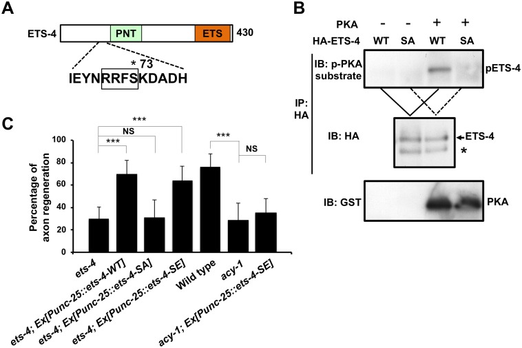
Fig 6. ETS-4 functions downstream of the cAMP pathway.
(A) A schematic diagram of ETS-4. The amino acid sequence around a PKA phosphorylation consensus site is shown below. The consensus site for PKA phosphorylation is boxed. The Ser-73 residue is indicated by an asterisk. (B) PKA phosphorylates ETS-4 at Ser-73 in vitro. In vitro phosphorylation of ETS-4 protein by PKA is shown in the lower panels. COS-7 cells were transfected with HA-ETS-4 (WT) or HA-ETS-4(S73A) (SA), and cell lysates were immunoprecipitated with anti-HA antibody. The immunoprecipitates were divided and then subjected to in vitro kinase assays using recombinant GST-fused active PKA. Phosphorylated ETS-4 was detected by immunoblotting with anti-phospho-PKA substrate rabbit monoclonal antibody. Asterisk indicates a heavy chain of IgG. (C) Percentages of axons that initiated regeneration 24 hr after laser surgery. Error bars indicate 95% SI. ***P<0.001; NS, not significant.

