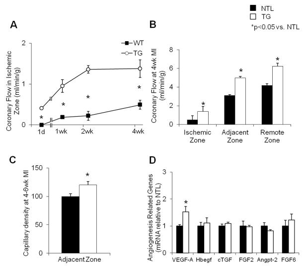
Fig. 3.
(A) Coronary blood flow in the ischemic zone at 1 day, 1 week, 2 weeks and 4 weeks after permanent CAO, showing that recovery of flow was significantly faster and of greater magnitude in TG vs. NTL rat hearts at the times indicated (n=4–6, p<0.05). (B) Coronary blood flow was increased in central ischemic, adjacent to ischemic and remote to ischemic zones of the heart. (C) Capillary density, normalized to NTLs, was increased in TG hearts at 4–6 wk post CAO in the zone adjacent to the scar. (D) Upregulation of angiogenic genes expressed in TG and NTL cardiomyocytes was determined by microarray analysis. The mRNA expression was further verified using qPCR. The significant increase in VEGF-A expression in TG hearts was verified. Results are expressed as the mean ± SEM. n=4–8; *p<0.05 vs. NTL. VEGF-A, vascular endothelial growth factor-A; Hbegf, Heparin-binding EGF-like growth factor; cTGF, connective tissue growth factor; FGF2, fibroblast growth factor-2; Angpt-2, angiopoietin 2; FGF6, fibroblast growth factor 6.

