Fig. 4.

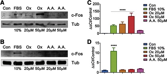
Oxalate induces expression of c-Fos in MCF-7 cells but not in HEK-293 cells. Cells were plated in six wells and grown to 80 % of confluence. Then cells were starved to achieve quiescence (see M & M). After that, each experimental condition was achieved by the stimulation with the specified reagent during 1.5 h. MCF-7 (a) or HEK-293 (b) cells were lysed, then the supernatant fractions were separated in 12 % SDS-PAGE gel and immunoblotted using anti-c-Fos antibody. α-Tubulin was used as loading control. c-fos expression was measured in MCF-7 (c) or HEK-293 (d) cells by qRT-PCR and normalized against housekeeping genes (GAPDH for MCF-7 cells and RPLPO for HEK-293 cells) using the Sequence Detection Software v1.4. Shown are the mean values of 3 independent determinations performed in quadruplicate. Con: Control, No reagent addition FBS: fetal bovine serum. Ox: oxalic acid. A.A.: acetic acid. Statistical significance determined by One- way ANOVA with Holm-Sidak’s test was performed in experiments shown in Fig. 4c and d, **** P value < 0.0001
