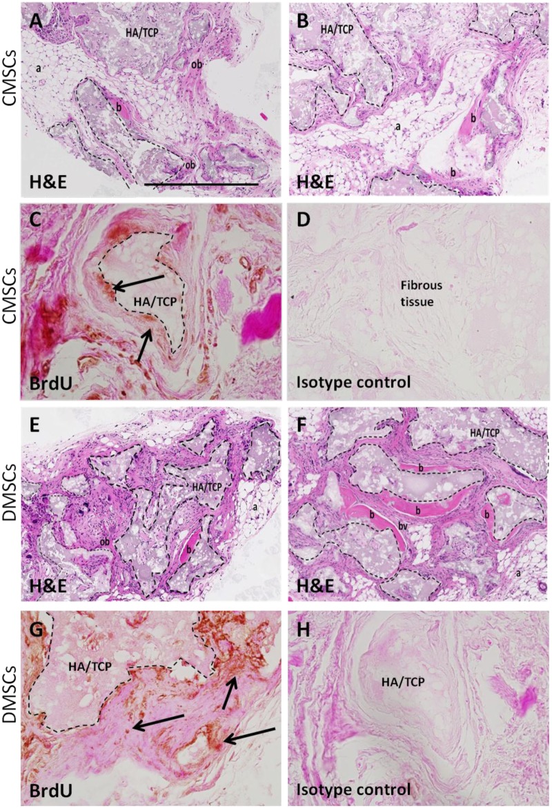
Fig 3. Histology of CMSCs and DMSCs transplants.
Cross sections are representative of CMSCs transplants (A-B) and DMSCs transplants (E-F) after 8 weeks stained with Haematoxylin and Eosin (H&E). In the transplant, the HA/TCP carrier surfaces (dashed lines) are lined with new bone formation (b), areas of immature bone (ob) together with the surrounding fibrous and hematopoietic tissue (a) and blood vessel (bv). Representative BrdU staining for localization of implanted CMSCs (C-D) and DMSCs (G-H). BrdU-stained implanted cells were found lining the mineralized matrix (black arrows) and surrounding fibrous tissue. Brown nuclear staining is indicative of DAB reactivity. There was no immunoreactivity present in sections stained with isotype-matched antibodies. HA/TCP: hydroxyapatite/tricalcium phosphate particles. Magnification is 100X and scalebar is 500 μm.

