
Fig. 1.
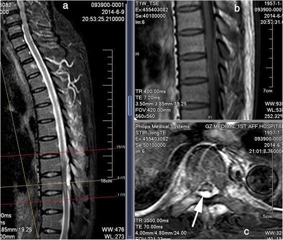
Magnetic resonance imaging of the thoracic vertebra. a Contrast-enhanced T2-weighted sagittal scan demonstrates a high-signal lesion behind the dural sac at the T8–9 level. b T1-weighted sagittal scan shows that the lesion has an area of moderate signal intensity and low-signal spots. c T2-weighted axial scan shows a high-signal area in both the vertebral canal and paravertebral muscle. The epidural abscess (single-headed arrow) is behand the spinal cord and dural sac