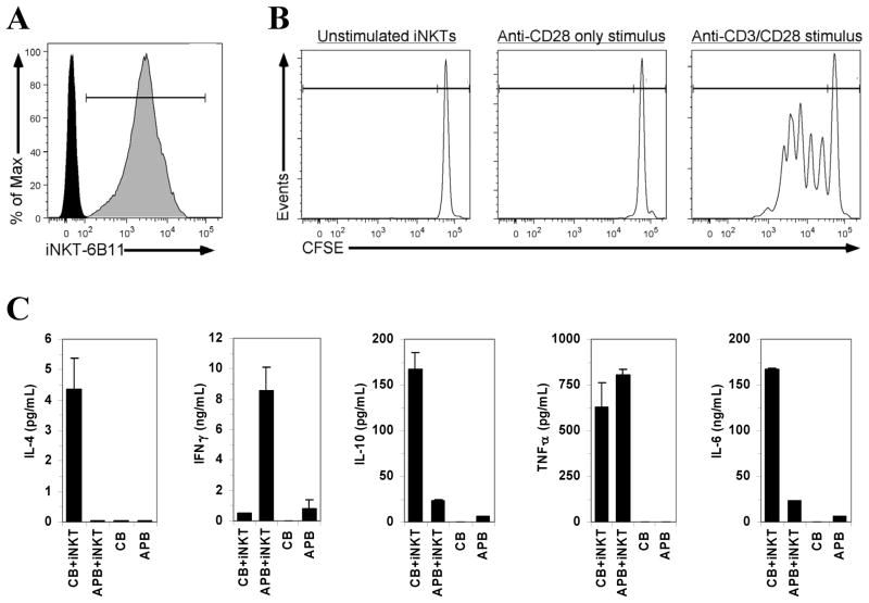
Figure 3. Characterization of purified human iNKT cells.
(A) Positive staining of CD1d tetramer-purified iNKT-cell with the 6B11 antibody. (B) iNKT cell proliferation when stimulated with plate-bound anti-CD3 (OKT3) and soluble anti-CD28 antibodies (as described in Ladd et al., 2010). (C) Cytokine profile of purified iNKT cells reconstituted (1 iNKT:1000 mononuclear cells) with CD1d-tetramer-depleted (FACS) autologous cord blood (CB) or adult peripheral blood (APB) mononuclear cells following stimulation with α-galactosylceramide (100 ng/mL; 96 hours). Cytokine levels are presented (mean, error bars: standard deviation of duplicate ELISA analyses) after subtraction of unstimulated cells.

