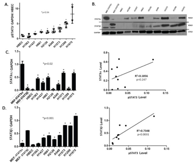
Figure 1. STAT3 is constitutively activated in most NSCLC cell lines, which correlates with levels of STAT3β.
A) Level of pSTAT3 were determined by Luminex beads in human NSCLC cell lines and in a normal human bronchial epithelial cell line (HBEC3-KT); values were corrected for GAPDH levels, expressed as a fraction of that obtained in HBEC3-KT cells, and the mean ± SD of 3 determinations shown; an asterisk (*) indicates those cell lines increased compared to HBEC3-KT cells (p<0.04). B) Levels of STAT3α, STAT3β and GAPDH were determined by immunoblot in lysates of murine embryonic fibroblast cells (MEF) lines in which the STAT3 gene was deleted followed by transient expression of STAT3α (MEF-STAT3α) or STAT3β (MEF-STAT3β)(13), HBEC3-KT cells, and human NSCLC cell lines (representative gel shown). Levels of STAT3α (C) and STAT3β (D) were quantified by densitometry using Image software. Each value was corrected using its corresponding GAPDH level and the mean ± SD of 2 determinations shown for each cell line (left panels) or as a function of its corresponding pSTAT3 level (right panels). An asterisk (*) indicates those cell lines increased compared to HBEC3-KT cells (p<0.02). pSTAT3 levels did not significantly correlate with levels of STAT3α (R2=0.1856, p=0.247), but did with levels STAT3β levels (R2=0.7348, p=0.0031).

