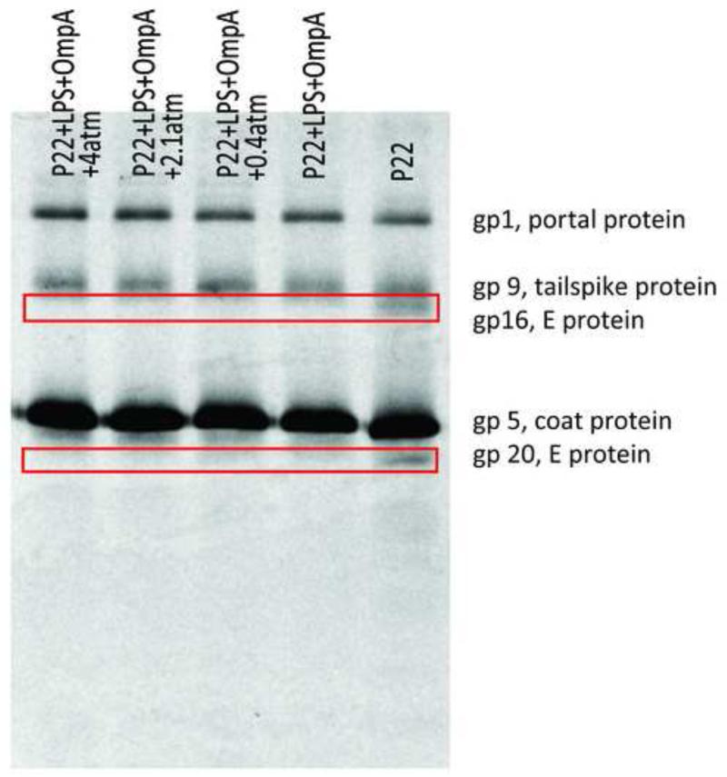
Figure 4. E proteins are ejected in the presence of both LPS and OmpA.
SDS-PAGE gel visualized by autoradiography showing the protein content in the pellet after ejection was triggered by LPS and OmpA at different external osmotic pressures. The right-most lane is the control which does not contain any receptor. This is a representative film that most clearly shows the release of gp 16 and gp 20.

