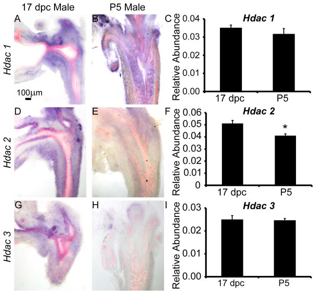
Figure 1. Type 1 histone deacetylase (Hdac) mRNAs are expressed and change in localization in fetal male mouse lower urinary tract during prostate budding and branching morphogenesis.
In situ hybridization (ISH, purple) for Hdac mRNAs combined with immunohistochemistry (IHC) using antibodies targeting epithelium (E-cadherin, red) were used to pinpoint localization of (A–B) Hdac1, (D–E) Hdac2 and (G–H) Hdac3 in male mouse lower urinary tract sections from 17 days post coitus (dpc) and postnatal day (P)5. QPCR was used to determine relative abundance of Hdac 1-3 mRNAs across whole male UGS at 17 dpc and P5 relative to a Ppi1 control (C,F,I). Results are mean ± SEM, n=5 mice per group. Asterisk indicates significant differences (p ≤ 0.01) between groups.

