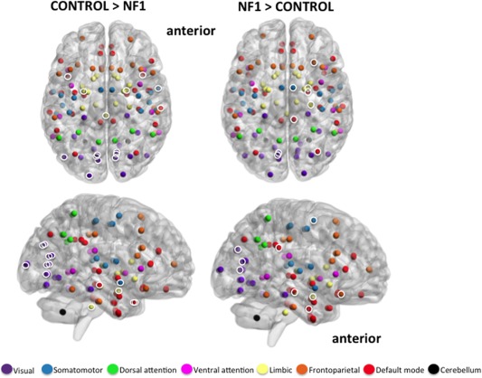
Figure 3.

Axial and medial view (right hemisphere) of significantly greater modular clustering in controls (left; 15 unique nodes) and NF1 participants (right; 9 unique nodes). Brain networks consist of several modules, or smaller “neighborhoods” of brain regions more connected to one another than to the rest of the network. “Modular clustering” evaluates the frequency with which any two nodes are members of the same module, and identifies nodes that are functionally related to one another. Highlighted nodes (white circles) are brain regions that consistently fall in the same module together, and do so significantly more often than identical nodes in the opposing group. Uncircled nodes have modular clustering patterns, but the pattern is not significantly different between groups. Results suggest that the largest difference between groups is in the clustering patterns of the visual (purple) and default mode (red) networks. Controls demonstrate a tightly clustered visual network and bilateral clustering of visual, default‐mode, and limbic nodes. NF1 participants show broader and more diffuse clustering patterns, combining nodes from visual, default mode, limbic, frontoparietal, and somatomotor networks. NF1 clustering is primarily in the right hemisphere. Anatomical locations of all 113 nodes are represented by spheres. Sphere colors match those specified in Figure 2. Module clusters shown at FDR 10%. [Color figure can be viewed in the online issue, which is available at http://wileyonlinelibrary.com.]
