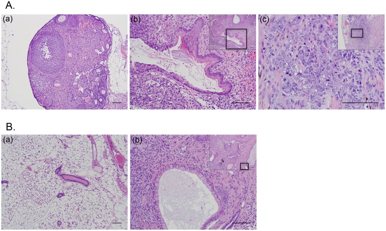
Fig 6. Histo-Pathological analysis of representative images (hematoxylin-eosin (H&E) tissue stain).
(A) Ovarian Panel: (a). Normal right (non-injected) ovary (100X). Scale bar is 100 μm; (b). Left Ovarian mass (following orthotopic inoculation with mESC) showing signs of a mature teratoma (small window, 100X) characterized with a focus of mature, keratinizing squamous epithelium within the area depicted inside the box (200X). Scale bar is 100 μm; (c). Ovarian mass (following orthotopic inoculation with mESC-HrasV12/SV40-LTg) showing signs of immature teratoma (small window, 100X) characterized with scattered foci of a high-grade malignant neoplasm within the area depicted inside the box (400X). Scale bar is 100 μm. (B) Breast Panel: (a). Normal murine mammary tissue (100X). Scale bar is 100 μm; (b). Breast mass (following orthotopic inoculation of mESC into cleared inguinal mammary fat pad) exhibiting signs of mature teratoma (small window, 100X) characterized with the respiratory-like epithelium having well formed cilia within the area depicted inside the box (200X). Scale bar is 100 μm.

