Fig. 4.

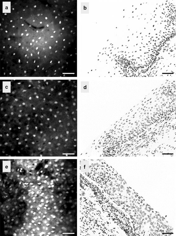
Confocal images of the cervix taken at a depth of 15 µm (left) and the corresponding histopathological section (right); a, b normal, c, d CIN1 lesion, and e, f CIN3 lesion. Scale bar is 50 µm

Confocal images of the cervix taken at a depth of 15 µm (left) and the corresponding histopathological section (right); a, b normal, c, d CIN1 lesion, and e, f CIN3 lesion. Scale bar is 50 µm