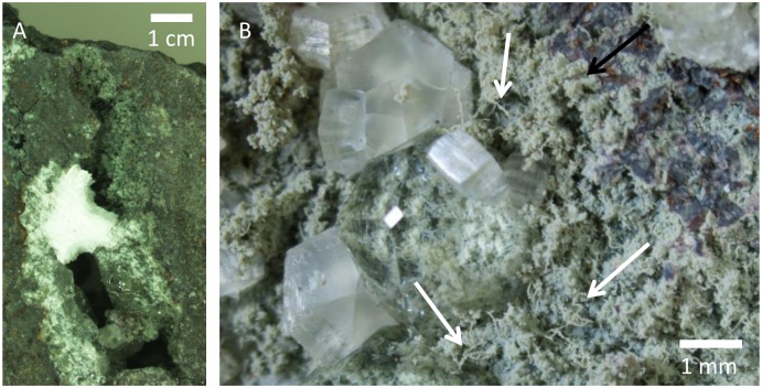
Fig 1. Overview of the vein.
Microphotographs of the fracture in sample (SMNH X5332). (A) Overview of the vein. (B) Showing parts of one side of the fracture after splitting. Dense fungal mycelia of yeast-like cells and hypha occasionally overgrown or intergrown by zeolites. Black arrow indicates the fungal mycelia preserved by montmorillonite, white arrows indicates specific hyphae.

