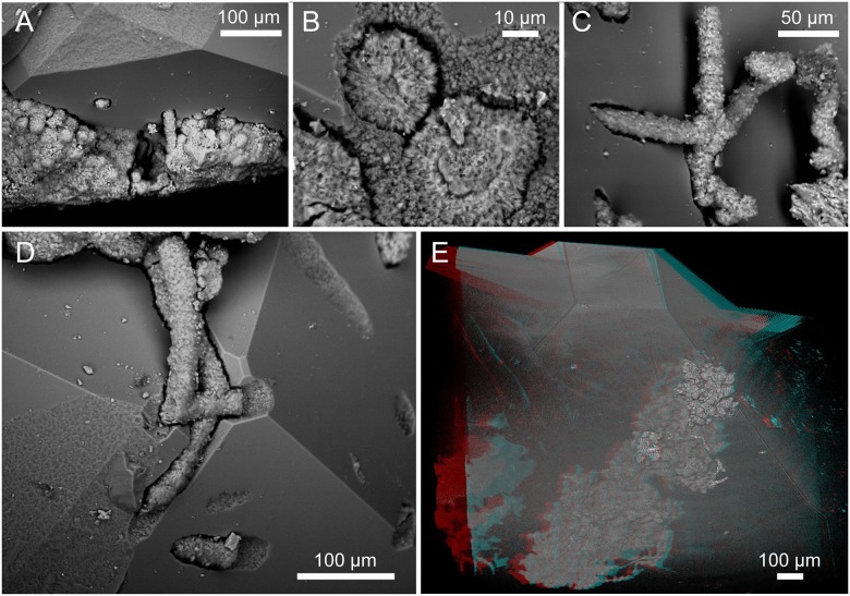
Fig 8. ESEM (A–D) (SMNH X5339) and SRXTM (E) (SMNH X5340) images of fungi influencing the mineral surface.
(A) The influence of both yeast and hyphae on the mineral surface leaving negative pits. (B) The contact between two protruding hyphae and the zeolite surface. Note the rough and irregular texture of the mineral surface at contact compared to the normally smooth surfaces. (C) Hyphae protruding angularly and creeping along the mineral surface. (D) Hyphae creep along the mineral surface and their influence on the zeolite surface at contact. (E) Stereo anaglyph of an assemblage of cells within a zeolite crystal.

