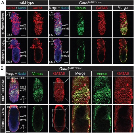
Fig. 4.

Co-expression of the Gata6 H2B-Venus reporter with endogenous GATA6 during early post-implantation development. a Wholemount immunofluorescence for endogenous GATA6 protein (red) on wild-type and Gata6 H2B-Venus/+ embryos at E5.5. Projected surface views showed that Venus and GATA6 were mostly co-localized in the VE. Corresponding projections showing internal views of the same embryos demonstrated that neither Venus nor GATA6 were expressed in the embryonic or extra-embryonic ectoderm at these stages. b Wholemount immunofluorescence for endogenous GATA6 protein (red) on Gata6 H2B-Venus/+ embryos at E6.0 (pre-streak, PS). The dashed white boxes indicate regions of higher magnification showing Venus and GATA6 expression in the VE. It should be noted that red fluorescent signal in the apical VE is likely due to non-specific binding of the GATA6 antibody to the surface of the extraembyonic tissue. Surface and internal views are renderings of z-series images. Extra-embryonic visceral endoderm (exVE), embryonic visceral endoderm (emVE), proximal (Pr), distal (D), anterior (A), posterior (P). Nuclei were stained with Hoechst (blue). Scale bars are 50 μm
