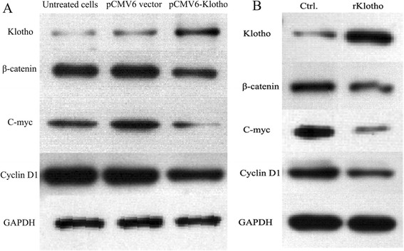
Fig. 5.

Klotho levels negatively correlate with Wnt/β-catenin signaling pathway in HepG2 cells. a The HepG2 cells with the density of 3 × 10⁴ cells/per well were plated in 24-well plates. Eight hours later, pCMV6-Klotho and p CMV6 vector were transfected into HepG2 cells. After 48 h, the expression of Klotho, β-catenin, C-myc, and Cyclin D1 was detected by western blotting. b The HepG2 cells with the density of 1 × 10³ cells/per well were plated in 24-well plates. Eight hours later, recombinated Klotho and BSA were added into HepG2 cells, respectively. After 48 h, the expression of Klotho, β-catenin, C-myc, and Cyclin D1 was detected by western blotting
