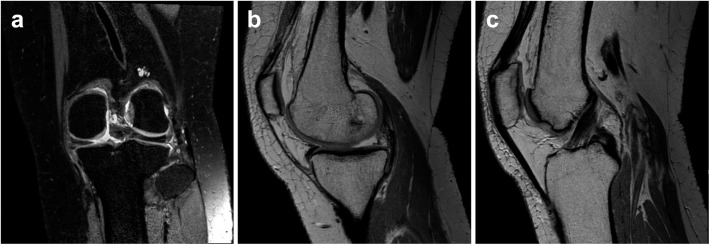
Fig 2. Knee MRI of a patient who had a medial meniscus root tear.

Fat-saturated proton-density-weighted images in the coronal scan (a) and proton-density-weighted sagittal (b) scan showed a defect in the medial meniscus posterior horn root. A medial extrusion of the meniscus and high-grade chondral lesions due to osteoarthritis are noted. A proton-density-weighted sagittal scan at a different level (c) showed an ACL that was intact.
