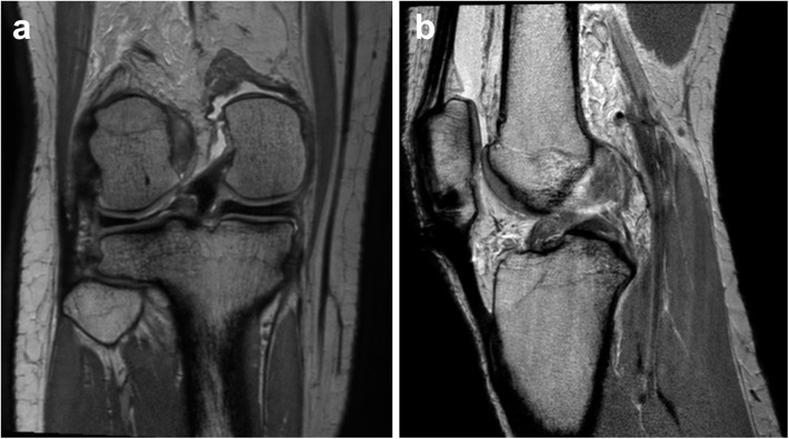
Fig 3. Knee MRI of a patient who had a lateral meniscus root tear.
Proton-density-weighted coronal scan (a) showed increased signal intensity on the lateral meniscal root, suggesting a meniscal tear. Although there was a meniscal root tear, meniscal extrusion was not evident. In addition, there was an anterior cruciate ligament tear. Focal discontinuity in the mid-segment of the anterior cruciate ligament with diffuse swelling was seen in the proton-density-weighted sagittal scan (b).

