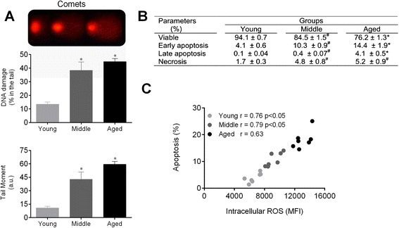
Fig. 3.

Augmented ROS leads to DNA damage and apoptosis of HSCs during aging. a The top panel shows typical photographs of comets with higher DNA fragmentation in the middle and aged groups. Bar graphs show the percentage of DNA in the tail and the tail moment. b Table shows the average percentage of viable cells, early apoptosis, late apoptosis and necrosis detected by annexin-V and PI staining. c Correlation between intracellular ROS levels and apoptosis. Values are means ± SEM, *p < 0.05 vs. young. # p < 0.05 vs. middle
