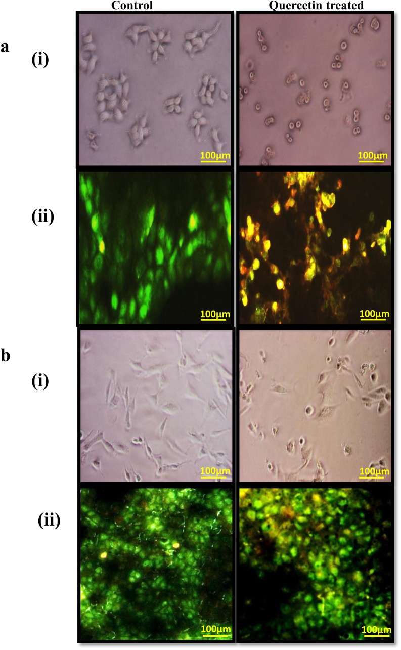
Fig 2. Cell pictures show the cell morphology and viability of MCF-7 and MDA-MB-231 control and quercetin treated cells.
a(i) MCF-7 cells cultured with and without quercetin for 24h were examined for changes in cell morphology and photographed using a phase-contrast microscope. a(ii) MCF-7 cells with and without quercetin for 24h were examined for the viability of cells by dual staining. b(i) MDA-MB-231 cells cultured with and without quercetin for 24h were examined for changes in cell morphology and photographed using a phase-contrast microscope. b(ii) MDA-MB-231 cells with and without quercetin for 24h were examined for the viability of cells by dual staining. The green stained are viable cells and the yellowish orange stained are damaged/apoptotic cells.

