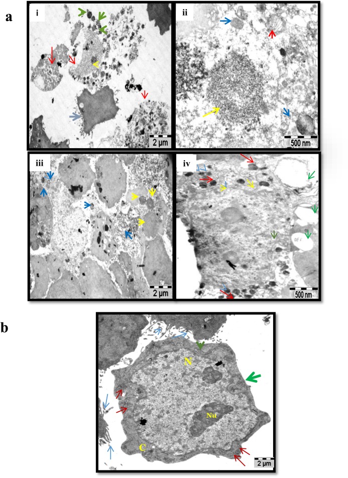
Fig 3. TEM picture represents the morphology and organelles of MCF-7 control and quercetin treated cells.
A. TEM analysis showing the structural changes and damages occur on treatment with quercetin. Picture (i) shows a undamaged cell (blue arrow) in the middle along with the cells undergoing apoptotic necrosis (red arrow), chromatin condensation inside the nucleus (yellow arrow) and autophagosomes (green arrow). Magnification at 500nm (ii) shows the condensation of the chromatin (yellow arrow), autophagosomes (blue arrow) and ER vesiculation (red arrow). (iii) Represents cells at lower magnification showing plenty of chromatin condensation (yellow) and autophagosis (blue) in group. (iv) Represents a single damaged cell showing huge number of autophagosis (red arrow), nuclear membrane showing starting of hetero chromatin (yellow), large number of vesicles in the cytosol (green arrow). B. The picture shows a single cell with microvilli (blue arrow), nucleus (N) present at the center of the cell, nucleoli (Nu), mitochondria (red arrow) (shows active energy production), structured Golgi apparatus (green arrow) and endoplasmic reticulum (orange arrow) finely organised. All these events represent a cell in active state.

