
Fig 1. Rosiglitazone-mediated sensitization of HCT116 cells to LA-12-induced apoptosis.
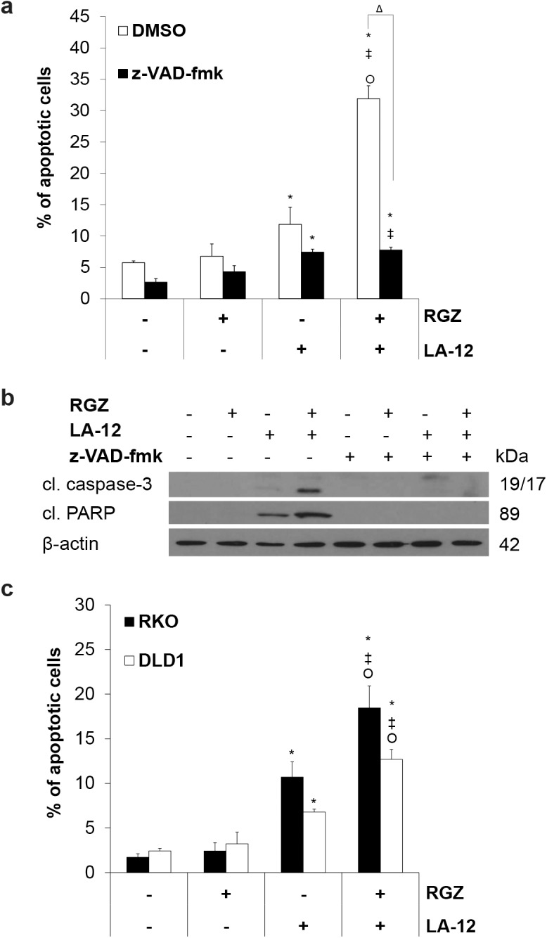
(a) Percentage of apoptotic (annexin V positive/propidium iodide negative, flow cytometry) HCT116 wt cells and (b) cleavage of caspase-3 and PARP (Western blotting) following pretreatment (24 h) with rosiglitazone (RGZ, 50 μM) and subsequent treatment (48 h) with LA-12 (0.75 μM), in the absence (DMSO) or presence of z-VAD-fmk (10 μM). (c) Percentage of apoptotic (annexin V positive/propidium iodide negative, flow cytometry) RKO or DLD1 cells following pretreatment (24 h) with rosiglitazone (RGZ, 50 μM) and subsequent treatment (48 h) with LA-12 (0.75 μM). Results are means + S.E.M. or representatives of three independent experiments. Statistical significance: P < 0.05, * versus control, ‡ versus RGZ, Ο versus LA-12, and Δ for with/without z-VAD-fmk.