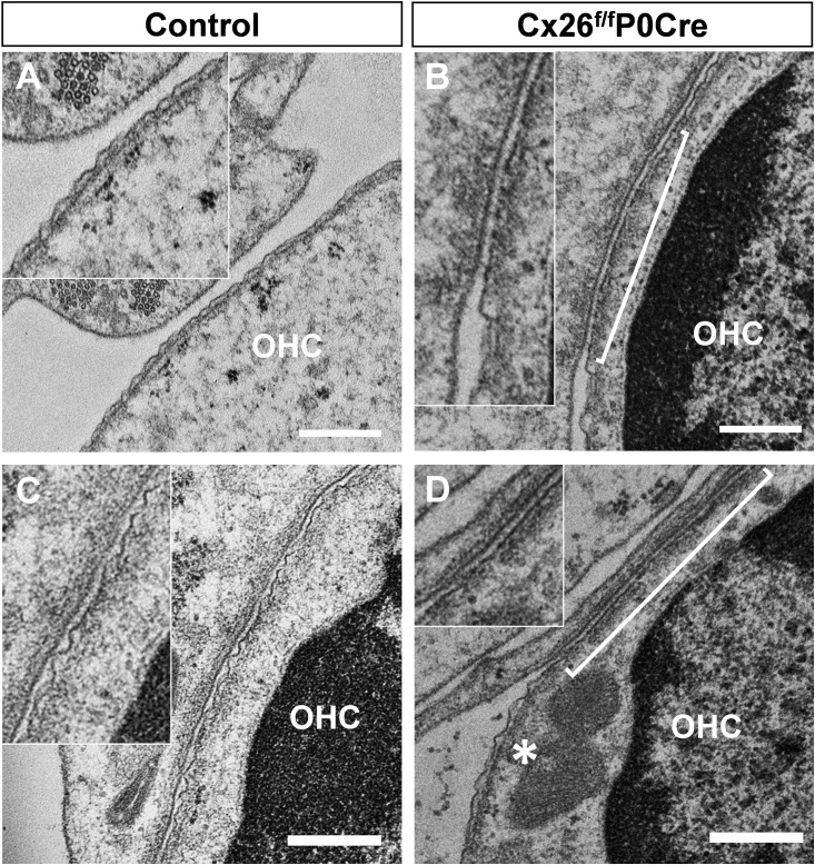
Fig 2. Ultrastructure of the plasma membrane of OHCs.
Transmission electron micrographs of OHCs. Ultrastructure of the plasma membrane of OHCs around apical part of cochlea in 5-week-old Cx26f/fP0Cre mice (B and D) and littermate controls (A and C). Horizontal ultrathin sections of the organ of Corti, including transverse section of OHCs, show the wavy surface of the plasma membrane that is thought to indicate the structure of the cortical lattice (A and C). In Cx26f/fP0Cre mice, a wavy surface structure was less apparent, and flat surfaces (brackets in B and D) were observed among the normal wavy surface regions of the plasma membrane (asterisk in D) at several points in OHCs. Scale bars, 500 nm. OHCs, outer hair cells.

