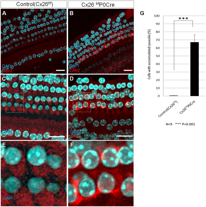
Fig 3. CAV2 accumulation in the organ of Corti.
(A-F) Immunofluorescence staining for CAV2 in the organ of Corti around apical part of cochlea in 3-week-old Cx26f/fP0Cre mice with GJB2-associated deafness and in littermate controls. Whole-mount cochleae were fixed and immunolabeled with anti-CAV2 (red). Nuclei were counterstained with DAPI (blue). In contrast to the controls, notable accumulation of CAV2 was observed at the organ of Corti in Cx26f/fP0Cre cochleae. (G) shows the mean percentage of cells with accumulated CAV2 in control and Cx26f/fP0Cre cochleae. There was a statistically significant difference between the control and Cx26f/fP0Cre mice. Values represent the mean ± SE (n = 5 mice). ***P = 3.72 × 10⁻⁵, Student’s t-test.

