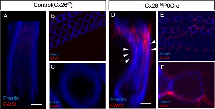
Fig 4. Subcellular localization of CAV2 in OHCs.
Accumulation of CAV2 was observed in OHCs around apical part of cochlea and colocalized with prestin in 3-week-old control and Cx26f/fP0Cre mice. z-stacks of images were collected at 0.5-μm intervals (B, C, E, F), and the images of OHCs were reconstructed with graphics software (A, D). Double labeling for CAV2 and prestin revealed that OHCs of Cx26f/fP0Cre mice had an altered, hourglass-like shape and that CAV2 accumulated near the basolateral plasma membranes. In contrast, CAV2 accumulation was mainly observed surrounding the shrunken site of OHCs in Cx26f/fP0Cre mice (arrowheads in D). OHCs, outer hair cells.

