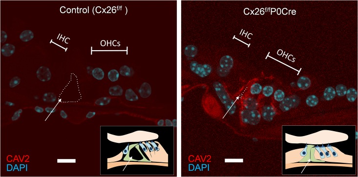
Fig 5. Immunolabeling of CAV2 in cryosections of the organ of Corti in 3-week-old control and Cx26f/fP0-Cre mice.
Cryosections of the organ of Corti around apical part of cochlea were immunolabeled with anti-CAV2 (red). Nuclei were counterstained with DAPI (blue). In contrast to the controls, the TC (dotted line with an arrow) was closed, and notable accumulation of CAVs was observed in OHCs and supporting cells. In particular, such accumulation was observed in cells surrounding the closed TC in Cx26f/fP0Cre cochleae. Scare bars, 10 μm. IHC, inner hair cell; OHCs, outer hair cells; TC, tunnel of Corti.

