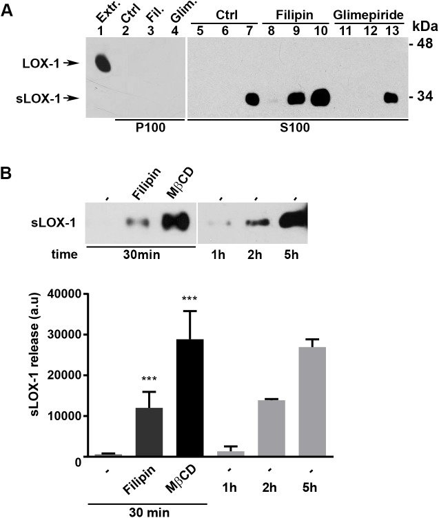
Fig 2. sLOX-1 release in COS cells.
(A) LOX-1-V5-COS transfected cells were treated with filipin (5μM) and glimepiride (5μM) for 15, 30 and 60 min. Conditioned media were centrifuged at 100,000 g and the resulting pellets (P100, lanes 2–4) and supernatants (S100, lanes 5–13) were analyzed by Western blotting. Lane 1 (Extr.) shows total protein extract (5 μg) loaded as a positive internal control of electrophoretic mobility of full-length LOX-1 receptor. (B) Upper panel shows sLOX-1 amount released in cells treated with Filipin (5μM) and MβCD (5mM) for 30 min compared to sLOX-1 constitutively released from untreated cells at 1, 2 and 5 hours. Lower panel shows the densitometric analysis for sLOX-1 band intensity. Data in histograms represent the average ± SEM of three experiments, P < 0.05 (*), P < 0.01 (**) or P < 0.001 (***).

