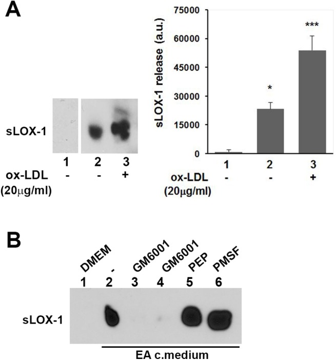
Fig 8. EA.hy926 cells secretome induce LOX-1 shedding.

(A) Western blot analysis of conditioned medium derived from cross-cells experiments. LOX-1-V5 COS transfected cells were incubated for 10 min with DMEM (lane 1), medium derived from untreated EA cells (lane 2) and medium derived from EA cells conditioned in the presence of ox-LDL (20μg/ml) (lane 3). The histogram on the right shows the densitometric analysis of sLOX-1 band intensity. (B) Effect of different classes of protease inhibitors on LOX-1 shedding. Inhibitors were added during the 10 min final incubation time. GM6001 was used at 2.6 and 26μM (lanes 3 and 4), Pepstatin A at 10μM (lane 5) and PMSF at 2mM (lane 6).
