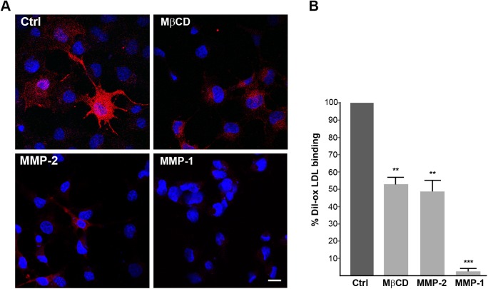
Fig 10. Soluble MMPs mediate LOX-1 shedding.
(A) Fluorescence analysis of LOX-1-V5 transiently transfected COS cells treated without (Ctrl) or 5 mM MβCD or 30μM active MMP-2 or active MMP-1 for 1 h at 37°C. Images show Dil-labelled ox-LDL (red fluorescence) for 1 h at 4°C. Nuclei are blue stained with Hoechst 33342. Scale bar 20 nm. (B) Histogram shows the percentage of red positive cells in different treatments, counting Hoescht-stained nuclei (n≥150). Data represent the average ± SEM of two different experiments.

