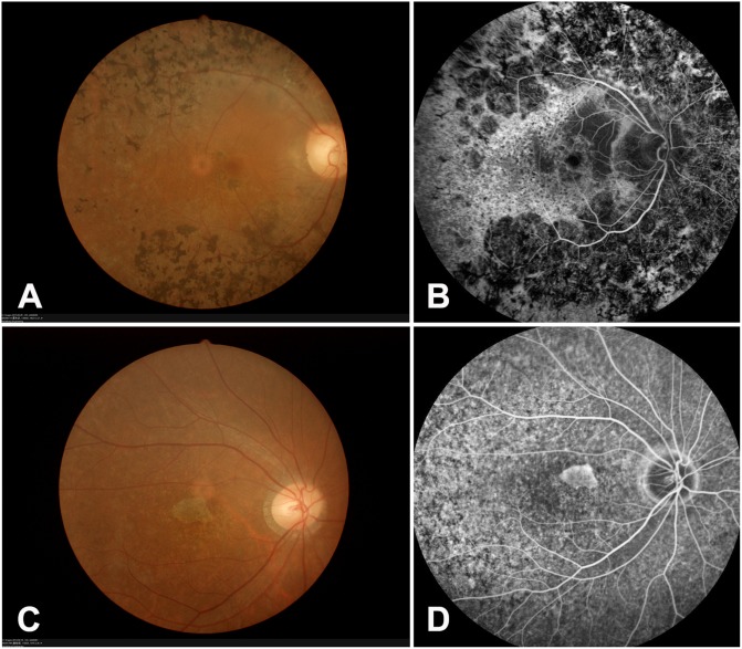
Fig 2. Representative fundus photography and fundus fluorescein angiography (FFA) images from RP98 and RP214 families.
(A-B) Proband of RP98. Fundus photography and fluorescein angiography (FFA) showing typical RP changes, including bone spicule-like pigmentation, retinal vascular attenuation, pallor of optic disc and chorioretinal degeneration. (C-D) Proband of RP214. Fundus photography and FFA showing attenuated arterioles and widespread RPE degeneration, the degenerated RPE area confused in the macular.

