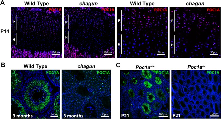
Fig 4. POC1A is expressed in the growth plate and seminiferous tubules.
A. POC1A immunostaining of sections from postnatal day 14 (P14) wild type and Poc1a cha/cha mutant tibial growth plates reveals strongest expression of POC1A (magenta) in the proliferative zone. B. POC1A immunostaining was carried out on testis sections from 3 mo old wild type and Poc1a cha/cha mutants. C. POC1A staining was also performed on testis sections from P21 wild type and Poc1a knockout mice. Scale bars as indicated.

