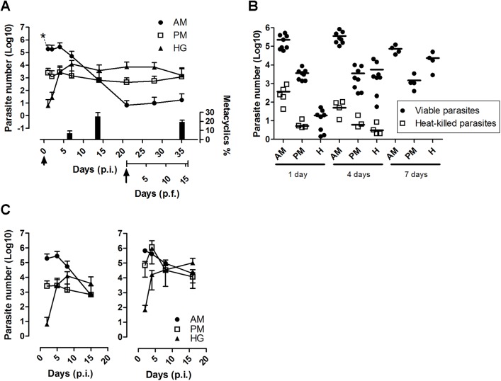
Fig 2. Real-time monitoring of T. cruzi loads in the different gut segments after R. prolixus infection.
(A) Follow-up of parasite development in the anterior midgut (AM), posterior midgut (PM) and hindgut (H) post-infection (p.i.) and post-feeding (p.f.). Adult insects were fed with 107 cells/ml of blood. The arrowhead and arrow indicates, respectively, the time of the initial infection and refeeding with a blood meal without parasites (21 days p.i.). Each time point represents three independent experiments (n≥8). The percentage of metacyclic trypomastigote forms (mean ± SE; n = 10) determined in the hindgut contents at 7 and 14 days p.i. and 14 days p.f. are indicated. (B) Monitoring of the DNA clearance of heat-killed parasites. Infections were performed with live parasites or parasites killed by incubation at 65°C during 2 hours before injection. n≥4 for each time point. (C) Comparison between the gut colonization of R. prolixus by T. cruzi Dm28c (left panel) and CL Brener (right panel). Each time point represents three independent experiments (n≥8). At several time points, the insects were dissected, and total DNA was extracted individually from the different gut segments and used to assess the parasite number by qPCR.

