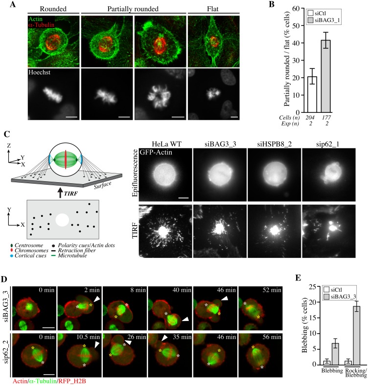
Fig 7. BAG3, HSPB8 and p62 regulate remodeling of mitotic F-actin structures.
(A) HeLa cells transfected with control siRNA or with BAG3-specific siRNA (siBAG3_1) were synchronized by the double Thymidine block method and processed for IF with anti-a-tubulin, phalloidin (F-actin) and Hoechst (DNA). Deconvolved confocal image stacks show F-actin and chromosomes in rounded mitotic cells compared to partially rounded and flat mitotic cells; Bars, 10 μm (B) Quantification of cells from A, indicating the proportion of siRNA-treated cells with defects in mitotic cell rounding; means +/- SD from 2 independent experiments. (C) Schemes depicting the proposed organization of retraction fibers in x, y, z view, with the deduced x, y view by TIRFM of actin dots connecting the mitotic cortex to the adhesive substrate. Representative TIRFM images from HeLa cells at metaphase transfected with the indicated siRNAs and transduced with BacMam-GFP-actin, showing a bright actin cortex sitting on the adhesive substrate surrounded by numerous actin dots in control cells, which are disorganized upon depletion of BAG3, HSPB8 or p62; the corresponding epifluorescence images are shown. Cells were imaged 48 h after transfection of siRNAs. (D) Confocal time-lapse sequences of Hela-RFP-H2B cells at metaphase that have been transfected with siBAG3_3 or sip62_1 and transduced with BacMam-GFP-a-tubulin and BacMam-RFP-actin, showing abnormal blebbing of the mitotic cortex designated by arrowheads which is associated with displacements of the spindle (S14, S15 and S16 Movies); asterisks designate spindle poles; Bars, 10 μm. (E) Quantification of cells from D, indicating the proportion of cells undergoing blebbing alone or blebbing with spindle rocking; means +/- SE from 158 (siCtl) or 195 cells (siBAG3) from 3 independent experiments (S1 Dataset).

