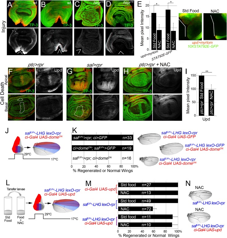
Fig 7. Cytokine signaling is controlled by ROS and JNK.
(A, B) In situ hybridization of upd (A) and upd3 (B) in wild type (wt) discs and JNKK hep r75 hemizygotes (C) after injury. (D) hep r75 hemizygote stained with anti-Upd after injury. (E) Mean pixel intensities of upd reporter (upd>myrtom Std food: 16.68 ± 3.22 and NAC: 9.01 ± 3.41, S.D.) and STAT92E reporter (10xSTAT92E-GFP Std food: 18.24 ± 2.73 and NAC: 8.63 ± 4.07, S.D.) after standard or NAC-supplemented food. White wedges indicate zone of injury. (F, G) Upd (anti-Upd) is mainly expressed in living cells and not in dead cells after ptc>rpr or sal>rpr. (H) Upd expression declines after NAC intake. TP-3: TP-PRO-3 nuclei staining. (I) Mean pixel intensities of Upd stained ptc>rpr discs with or without NAC feeding (150.29 ± 7.11 and 96.69 ± 18.97, S.D.). (J) Inhibition of the JAK/STAT signaling within dome DN impairs wing regeneration. Genetic design (J) using double transactivator system (as in Fig 2) to induce death (blue) and activate dome DN (red). (K) Percentage of regenerated wings for controls (rpr or dome DN expression only) and experimental (rpr and dome DN dual expression). Note that dome DN wings were not able to regenerate (rpr and dome DN dual expression), whereas dome DN wings in the absence of cell death are normal. Examples of wings (left) of controls and experimental. (L) Experimental design for testing the rescue of NAC effects by ectopic activation of upd. (M) NAC effect on repair ability was rescued by upd overexpression. Quantification of the percentage of wings that regenerate after NAC feeding for the indicated genotypes. (N) Examples of wings from NAC-feeding with rpr-ablation defects (upper) and with rescue after rpr-ablation and upd activation (lower). *P<0.05 **P<0.01.

