Figure 2.
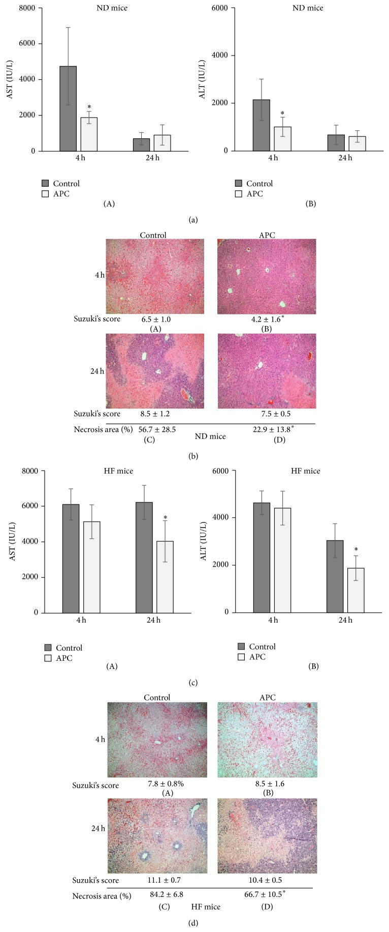
Transaminase levels and histology in ND and HF mice. Serum AST and ALT levels at 4 h were significantly decreased in ND-APC compared with ND-Control mice (∗ P < 0.05). There was no significant difference at 24 h between ND-APC and ND-Control mice ((a)-(A), (a)-(B)). H&E staining showed that the ND-APC had significantly preserved lobular architecture and reduced intrasinusoidal/vascular congestion compared with ND-Control at 4 h ((b)-(A), (b)-(B)). The necrotic area within livers was significantly reduced in ND-APC compared with ND-Control mice at 24 h (∗ P < 0.05) ((b)-(C), (b)-(D)). While serum AST and ALT levels at 24 h were significantly decreased in HF-APC compared with HF-Control mice (∗ P < 0.05), there was no significant difference at 4 h between HF-APC and HF-Control mice ((c)-(A), (c)-(B)). In HF mice, however, liver tissues in both the HF-APC and HF-Control groups showed marked changes in vacuolization and intrasinusoidal/vascular congestion ((d)-(A), (d)-(B)). Although there was more severe necrosis in HF mice than in ND mice, the necrotic area of hepatocytes was significantly reduced in HF-APC compared with HF-Control mice at 24 h (∗ P < 0.05) ((d)-(C), (d)-(D)). The numbers under the pictures show the modified Suzuki score and the percentage of necrotic area (%). The original magnification was ×100 (b, d).

