Figure 3.
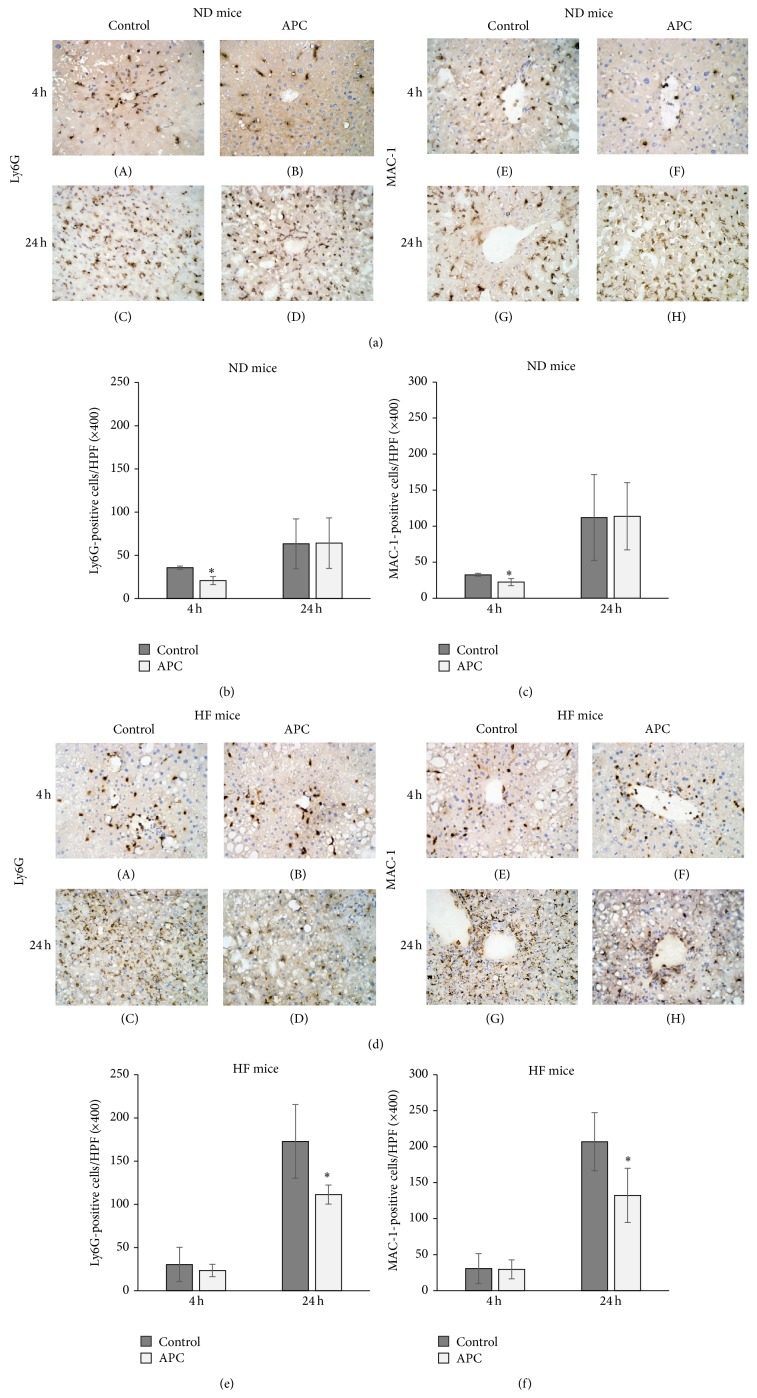
Leukocyte infiltration 4 h and 24 h after reperfusion. Infiltration of Ly6G-positive cells ((a)-(A), (a)-(B), and (b)) and MAC-1-positive cells ((a)-(E), (a)-(F), and (c)) in the liver was significantly decreased in ND-APC compared with ND-Control mice at 4 h (∗ P < 0.05). However, in HF mice, infiltration of Ly6G-positive cells ((d)-(C), (d)-(D), and (e)) and MAC-1-positive cells ((d)-(G), (d)-(H), and (f)) was significantly decreased in HF-APC livers compared with HF-Control livers at 24 h (∗ P < 0.05). There was no significant difference at 4 h between HF-APC and HF-Control mice. The original magnification was ×400 (a, d).

