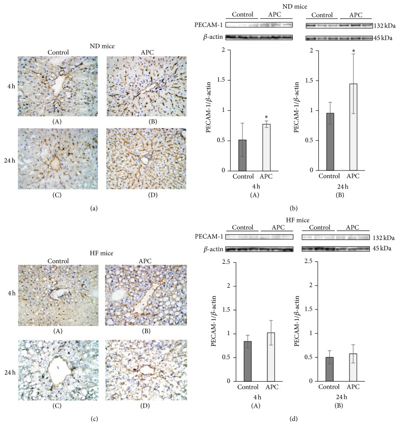
Figure 4.
Sinusoidal endothelial cell damage in ND and HF mice. IRI disrupted sinusoidal vasculature regardless of APC administration in HF mice. In the liver tissue of ND mice, there was significant large number of sinusoidal endothelial cells, which were stained with PECAM-1 antibody in ND-APC than in ND-Control mice at 4 h ((a)-(A), (a)-(B), and (b)-(A)) and 24 h ((a)-(C), (a)-(D), and (b)-(B)). In liver tissue of HF mice, sinusoidal endothelial cells which were stained with PECAM-1 antibody were disrupted regardless of APC administration at both 4 h ((c)-(A), (c)-(B), and (d)-(A)) and 24 h ((c)-(C), (c)-(D), and (d)-(B)).

