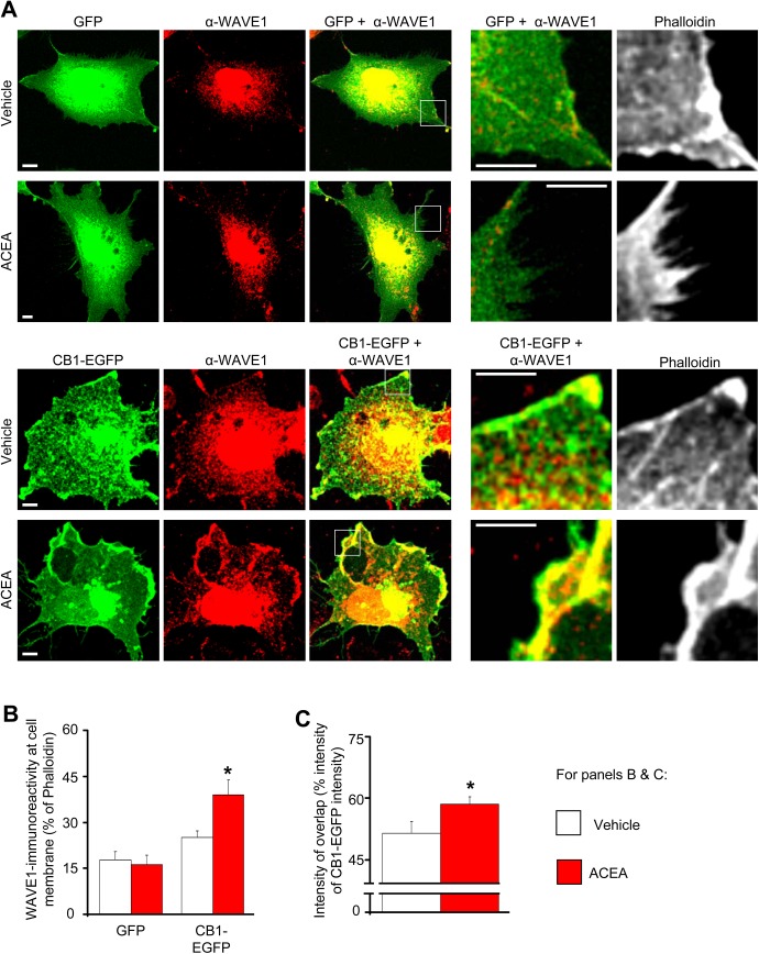
Fig 2. Increased localization of colocalization of WAVE1 and CB1-EGFP at the cell membrane in heterologously-transfected COS7 cells upon ACEA treatment.
(A) Distribution of heterologously-transfected WAVE1 in cells cotransfected with either GFP (control, upper panels) or CB1-EGFP (lower panels) following treatment with vehicle (DMSO 1:30,000) or CB1 agonist (ACEA 100 nM) for 45 min. Scale bar represents 5 μm. (B) Quantitative summary of intensity of WAVE1-immunoreactivity relative to Phalloidin-stained actin at the plasma membrane in GFP- or CB1-EGFP-coexpressing COS7 cells treated with vehicle (white bars) or ACEA (red bars). (C) Quantitative summary of CB1-EGFP-WAVE1 colocalization in vehicle- or ACEA-treated cells calculated as a fraction of the total intensity of CB1-EGFP fluorescence. Values in panels B and C represent the mean ± SEM and are derived from analyses on at least 15 COS7 cells each over several independent culture experiments. *p < 0.05 one way ANOVA followed by posthoc Tukey’s test.

