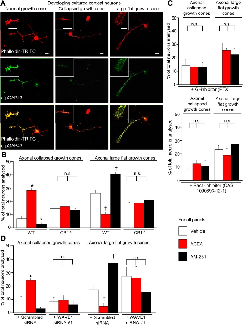
Fig 5. Cannabinoids regulate growth cone morphology via CB1-Gi-Rac1-WAVE1 signaling.
(A) Characterization of changes in growth cone morphology in developing cultured cortical neurons immunostained with anti-pGAP43 to recognize normal or enlarging growth cones and Phalloidin to label actin. Insets show normal, enlarged, or collapsed morphology in developing cortical neurons; scale bar represents 10 μm in all images and insets. (B) Bidirectional modulation of the proportion of growth cones with collapsed or enlarged morphology upon treatment with ACEA (100 nM), AM251 (600 nM) or vehicle (DMSO 1:30,000) for 1 h in neurons derived from wild-type mice (n = 10 independent culture experiments), but not in neurons from CB1-/- mice (n = 4). (C–F) Lack of modulation of cannabinoidergic modulation of growth cone morphology upon treatment in the presence of PTX (100 ng/ml), Rac1 inhibitor, CAS 1090893-12-1 (50 μM) or in neurons with siRNA-mediated downregulation of WAVE1; control siRNA-transfected neurons showed significant cannabinoidergic modulation (n = 4). All graphs represent mean values ± SEM. *p < 0.05, one-way ANOVA followed by posthoc Tukey’s test. N.s. stands for not significant.

