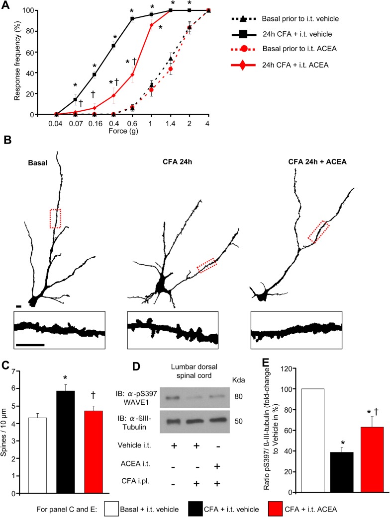
Fig 8. Intrathecal delivery of ACEA inhibits mechanical nociceptive hypersensitivity and nociceptive activity-induced structural plasticity of dendrites on spinal dorsal horn neurons in inflammatory pain conditions in vivo.
(A) Frequency of paw withdrawal responses to mechanical force via plantar application of graded von Frey hairs recorded prior to and 24 h after hind paw intraplantar injection of CFA. Leftward shift in response curves following CFA (indicative of hypersenstivity) is diminished in mice treated intrathecally with ACEA (2 pmol) over 24 h as compared to mice intrathecally receiving vehicle (n = at least 8 per group). (B) Traced images of Golgi-stained large pyramidal-like neurons in laminae II and V of the spinal dorsal horn and dendritic spines (insets) in mice represented in (A). Scale bars represent 10 μm in all panels. (C) Quantitative summary of dendritic spine density in spinal neurons from mice represented in (A) and (B); CFA-induced enhancement in spine density does not occur in mice receiving intrathecal ACEA (n = 12–16 neurons counted from over 4 mice per group). (D, E) Mice with hind paw inflammation show reduced levels of pSer397 WAVE1, quantified over βIII-tubulin as loading control, in spinal lumbar segments L3-L5 at 24 h post-CFA, which is partially and significantly reversed by intrathecal ACEA as compared to vehicle. An example of western blot analysis and quantitative summary from seven mice per group is shown in (D) and (E), respectively. All graphs represent mean values ± SEM *p < 0.05 as compared to basal values within the group and †p < 0.05 as compared to corresponding values in the vehicle group, two-way (A) and one-way (C, E) ANOVA for repeated measures followed by posthoc Tukey’s test.

