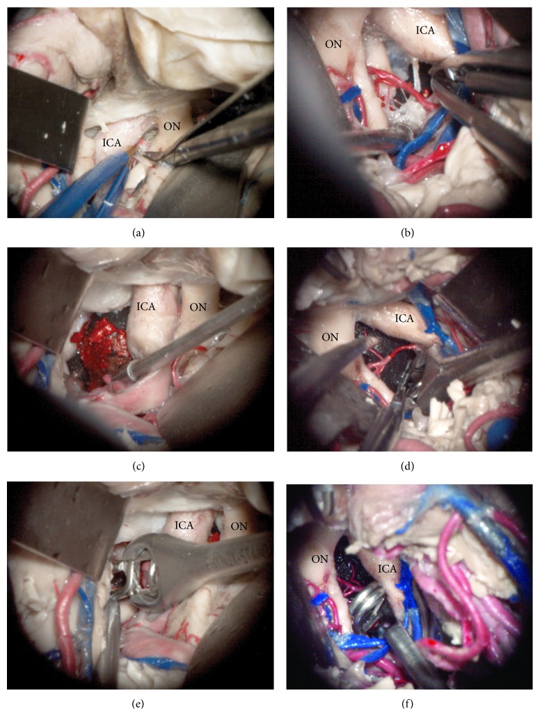
Figure 3.
Microscopic photography of left (a, c, and e) and right (b, d, and f) surgical simulation experiments for implanting and clipping a basilar tip aneurysm. After a wide sylvian fissure split, the left carotid cistern was dissected to access the interpeduncular fossa (a). The 3D aneurysm model was successfully implanted through the oculomotor triangle and positioned to match the target anatomy, basilar apex (c). Next, an angled aneurysm clip was introduced and positioned across the aneurysm neck in the basilar apex (e). Then, the right transsylvian approach was performed with the aneurysm model already implanted. The arachnoid dissection and perforator arteries manipulation were affected by the aneurysm mass effect (b). The dome of the previously implanted aneurysm was mobilized to identify the aneurysm neck (d) and an angled clip was applied to the basilar apex aneurysm neck (f). ICA: internal carotid artery, ON: optic nerve.

