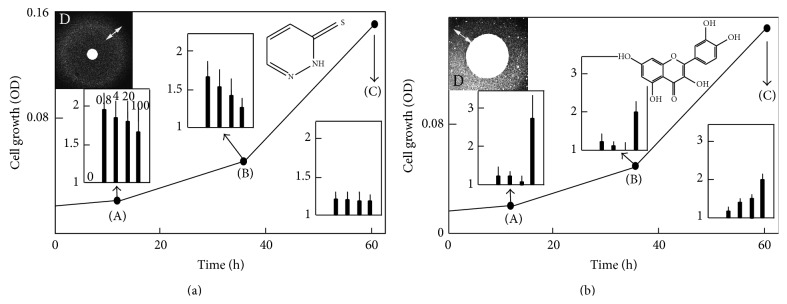
Figure 2.

Effect of drugs on the kinetics of Δyfh1 cell growth. Panels A (effect of LPS-01-04-L-G10) and B (effect of LPGS-02-C06) show a typical growth curve of Δyfh1 cells in liquid YNB-Raf medium with no addition as a reference curve and in inserts, the effect of the drugs on the kinetics of growth evaluated by measuring the cell density (OD600 nm) at 3 different stages of the growth: 12 h (A), 36 h (B), and 60 h (C) after addition of the drug at different concentrations (0.8, 4, 20, and 100 μM). The values in insert graphs represent the increase of growth due to the drug at these various concentrations, as n-fold increase of the cell density compared to the DMSO control (1 = no change, 2 = 2-fold increase, etc.). Dose-dependent effect is also illustrated by agar disc diffusion assays (YNB-Raf/agar medium) in panels D: paper discs (diameter of 0.3 cm) were impregnated with 7 μL of the concentrated drugs (10 mM in DMSO), and the pattern of growth of Δyfh1 colonies around the discs was photographed after 2–4 days. The zones showing the highest density of colonies are indicated by a double arrow. All of the experiments were performed in quadruplicate.
