
Fig. 2.
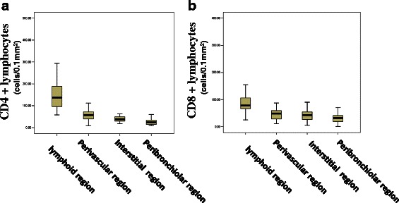
a Distribution of CD4+ T lymphocytes in different regions of lung tissue. The different in CD4+ cell counts between any two regions were statistically significant (p < 0.001). b Distribution of CD8 + T lymphocytes in different regions of lung tissue. CD8+ cell in follicles were much more than those in each of the other three regions (p < 0.001)