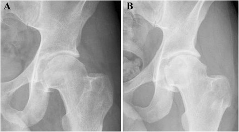
Fig. 2.

Initial and follow-up radiographs of the left hip of a 31 year-old male with acetabular osteoid osteoma. a Plain x-ray of the left hip before undergoing radiofrequency ablation or hip arthroscopy showing mild degenerative changes of the hip joint; b Plain x-ray of the left hip of the same patient 44 months following hip arthroscopy and radiofrequency ablation of the acetabular osteoid osteoma not showing further progression of the osteoarthritic changes
