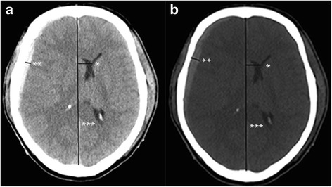
Fig. 1.

Depicting a CT scan of a patient who suffered from a right sided acute subdural hematoma. The standard windows W/L is shown in (a), and also the method to measure the thickness of the hematoma (**) and the midlineshift (midline ***, shift *). The thickness of the hematoma was 5 mm and the MLS 15 mm. After adapting the windows W/L was to the suggested level (b) the thickness of the hematoma was 10 mm and the MLS 15 mm
