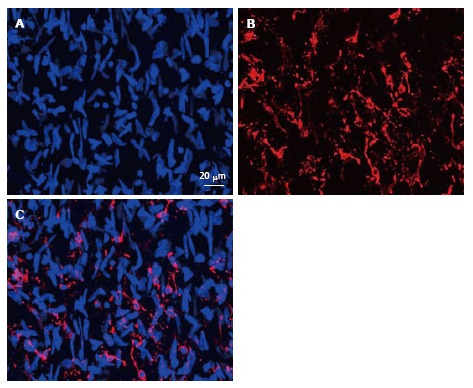
Figure 4.

Immunofluorescent images representing the merge of 45 optic “confocal” sections of the intima obtained with interval of 1 μm between consecutive sections (A and B). A: Nuclei were visualized by staining with ethidium bromide; B: Distribution and networks formed by HLA-DR+ cells, detected with the use of anti-HLA-DR antibody conjugated with Alexa 633; C: A merge of the images shown in (A) and (B). Human aorta specimen. Reproduced from Bobryshev et al[74], with permission from Elsevier. HLA: Human leukocyte antigen.
