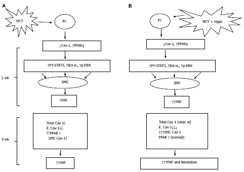
Figure 10.

Monocrotaline injury to endothelial cells resulting in the loss of caveolin-1 and the activation of the proliferative pathways (PY-STAT3, Bcl-xL, p-ERK) leading to PH at 2 wk; and a reciprocal relationship between caveolin-1 and peroxisome proliferator-activated receptorγ expression (A) and MCT + hypoxia (MCT + Hypo) accelerates the disease process (B). At 4 wk, there is a further loss of endothelial caveolin-1 (E. Cav-1) and enhanced expression of cav-1 in smooth muscle cells (SMC), however, the total cav-1 levels remain low (17% vs C, 100%). These alterations are accompanied by a further increase in pulmonary artery pressure. Panel B shows MCT + hypoxia (MCT + Hypo) accelerates the disease process. At 2 wk, extensive endothelial caveolin-1 is accompanied by the activation of proliferative pathways and PH (higher pulmonary artery pressure compared with MCT alone group). At 4 wk, a further loss of E. Cav-1 is accompanied by significantly increased expression of caveolin-1 in SMC compared with MCT alone group. At this stage total caveolin-1 level is closer to normal (81% vs C, 100%), and neointimal lesions can be seen. MCT: Monocrotaline; EC: Endothelial cells; PPAR: Peroxisome proliferator-activated receptor.
