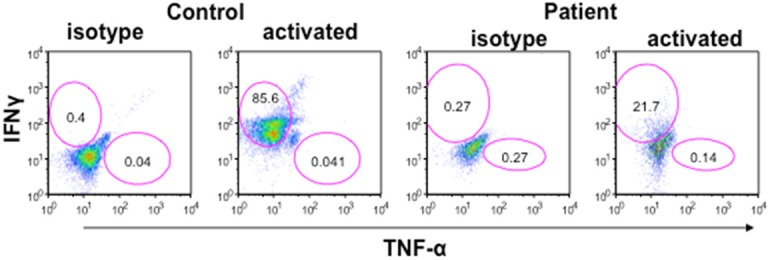
Figure 2.

IFN-γ and TNF-α containing CD4+ (Th1) cells. Mononuclear cells were stimulated with PMA and Ionomycin and secretion of cytokine was blocked by brefelidine. Cells were stained for surface CD4, fixed and then stained for Intracellular IFN-γ and TNF-α with respective antibodies and isotype controls. Cells were gated on CD4+ T cells and then analyzed for IFN-γ+ and TNF-α+ cells by multicolor flow cytometry. IFN-γ+ cells were markedly decreased (21.7%) as compared to control (85.6%). TNF-α+ cells were comparable. Blue lines are for isotype control and red lines for specific antibodies.
