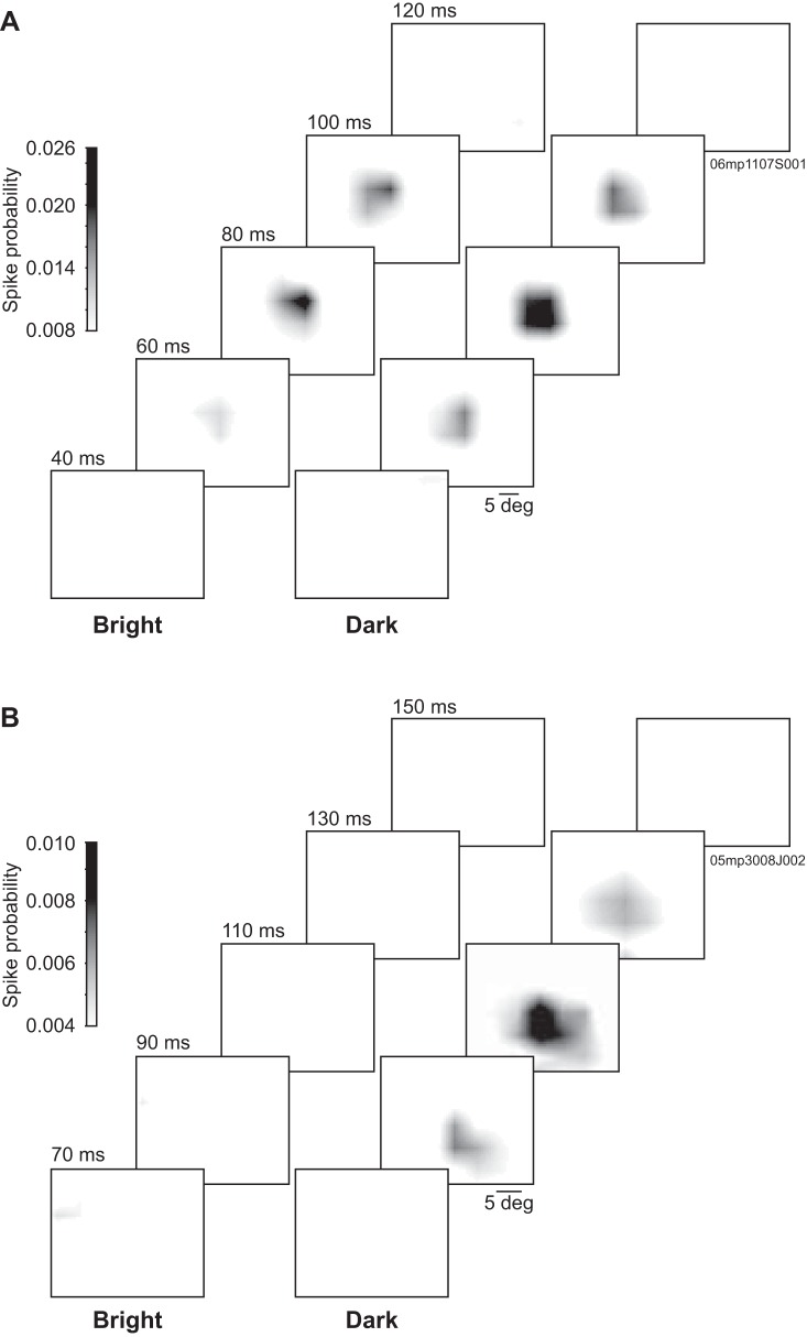
Fig. 1.
Representative spatiotemporal receptive field (RF) profiles from lateral posterior nucleus (LP) neurons. Grayscale-coded probability spatial maps from LP neurons obtained at different prespike times shown separately for bright and dark stimuli. A: this lateral LP (LPl) neuron responded to both bright and dark stimuli with spatially overlapping RF subfields and with coinciding peak probability latencies of ∼80 ms. This neuron was probed with 6.5° squares distributed on a 15 × 11 stimulation position grid. B: this medial LP (LPm) neuron responded to dark stimuli only, with a peak probability latency of ∼110 ms. This neuron was probed with 5° squares distributed on a 20 × 15 stimulation position grid.

