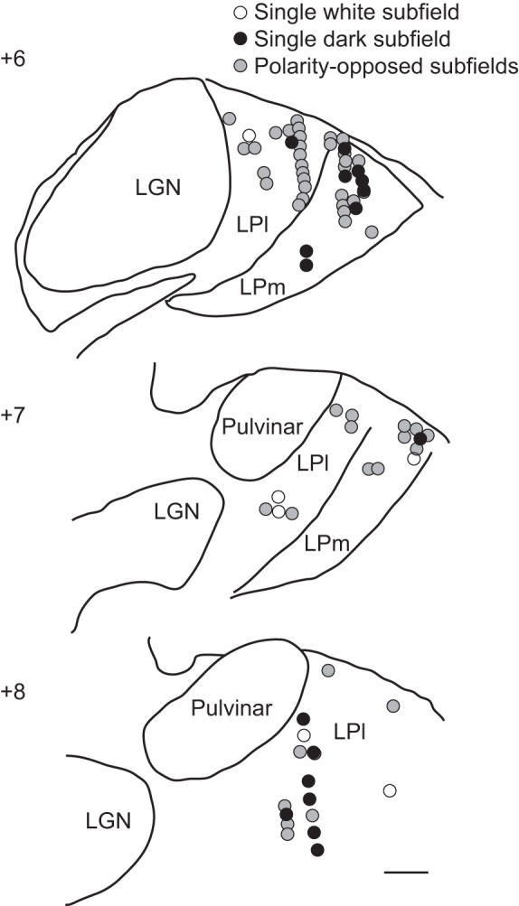
Fig. 9.

Anatomic distribution of recording sites. Schematic drawings of thalamic coronal sections at different antero-posterior coordinates indicating the anatomic position of the neurons studied. Neurons with polarity-opposed subfields are indicated by gray circles, whereas neurons with single bright or dark subfields are indicated by open and filled circles, respectively. LGN, lateral geniculate. Scale bar is 1 mm.
